




The word “cancer” often brings a wave of anxiety, but in the modern medical landscape, knowledge truly is power. Among the many questions women ask about their health, one of the most frequent is: Can ultrasound detect breast cancer early?
As breast cancer awareness grows, more women are seeking proactive ways to monitor their health. Early detection remains the single most important factor in surviving breast cancer, often making the difference between a simple, minimally invasive procedure and a long, difficult treatment journey. While mammography has long been the “gold standard” for screening, breast ultrasound has emerged as a vital, life-saving tool—particularly for women with dense breast tissue or those who are younger.
Breast ultrasound can help detect breast abnormalities, especially in women with dense breast tissue. While it may identify suspicious masses and cysts, it is often used alongside mammography and clinical evaluation for accurate breast cancer diagnosis and early-stage detection when tumors are too small to be felt.
Introduction: The Shift Toward Proactive Breast Health
Breast cancer is one of the most common cancers affecting women worldwide. However, the narrative around the disease is shifting from one of fear to one of empowerment through early detection. For decades, the mammogram was the only household name in screening. Today, we understand that a “one size fits all” approach doesn’t work for every woman.
Many women wonder if an ultrasound is enough to catch cancer in its infancy. They might have heard that ultrasound is “better” for dense breasts or that it avoids the radiation associated with X-rays. In this comprehensive guide, we will explore the nuances of breast ultrasound, how it compares to other imaging techniques, and why it is a critical component of early diagnosis.
Understanding Breast Cancer and the Importance of Early Detection
How Breast Cancer Develops
Breast cancer begins when cells in the breast start to grow out of control. These cells usually form a tumor that can often be seen on an X-ray or felt as a lump. The cancer becomes “malignant” (cancerous) if the cells can grow into surrounding tissues or spread (metastasize) to distant areas of the body.
Why Early Detection Improves Outcomes
When breast cancer is detected early—specifically in Stage 0 or Stage 1—the cancer is confined to a very small area. In these early stages, the survival rate is exceptionally high (often above 95–99%). Detecting a tumor when it is only a few millimeters in size, long before a woman or her doctor can feel it during a physical exam, is the ultimate goal of screening.
Survival Benefits of Early Diagnosis
Early diagnosis allows for:
-
More Treatment Options: Patients may qualify for breast conservation surgery (lumpectomy) rather than a mastectomy.
-
Less Aggressive Treatment: There is a higher likelihood of avoiding intensive chemotherapy if the cancer is caught before it spreads to lymph nodes.
-
Higher Quality of Life: Shorter treatment durations mean a faster return to normalcy.
Screening vs. Diagnosis
It is important to distinguish between these two terms.
-
Screening is testing done on people who have no symptoms (like a routine yearly mammogram).
-
Diagnosis is testing done to investigate a specific symptom (like a lump) or an abnormal result found during screening. Ultrasound plays a massive role in both, but it is particularly dominant in the diagnostic phase.
What Exactly Is a Breast Ultrasound?
A breast ultrasound, also known as sonography, is a non-invasive imaging test that creates a picture of the internal structures of the breast.
How Ultrasound Technology Works
Unlike mammograms, which use low-dose ionizing radiation (X-rays), ultrasound uses high-frequency sound waves. A handheld device called a transducer is moved over the skin. It sends sound waves into the breast tissue, which bounce off solid structures and fluids, creating “echoes.” These echoes are converted in real-time into images on a monitor.
Sound Waves vs. Radiation
Because ultrasound uses sound waves, it is 100% radiation-free. This makes it a preferred method for:
-
Pregnant women.
-
Women under the age of 30.
-
Women who require frequent follow-up monitoring.
Types of Breast Ultrasound
-
Targeted Ultrasound: Focuses on a specific area where a lump was felt or an abnormality was seen on a mammogram.
-
Whole-Breast Ultrasound (ABUS): An automated system that scans the entire breast, often used as a screening supplement for women with dense tissue.
When Doctors Recommend Breast Ultrasound
A Breast Cancer Specialist in Lucknow may recommend an ultrasound if:
-
You have dense breast tissue.
-
A mammogram shows a suspicious area that needs a closer look.
-
You are young (under 35) and have a palpable breast lump.
-
You are pregnant and have breast concerns.
-
A doctor needs to guide a needle during a biopsy.
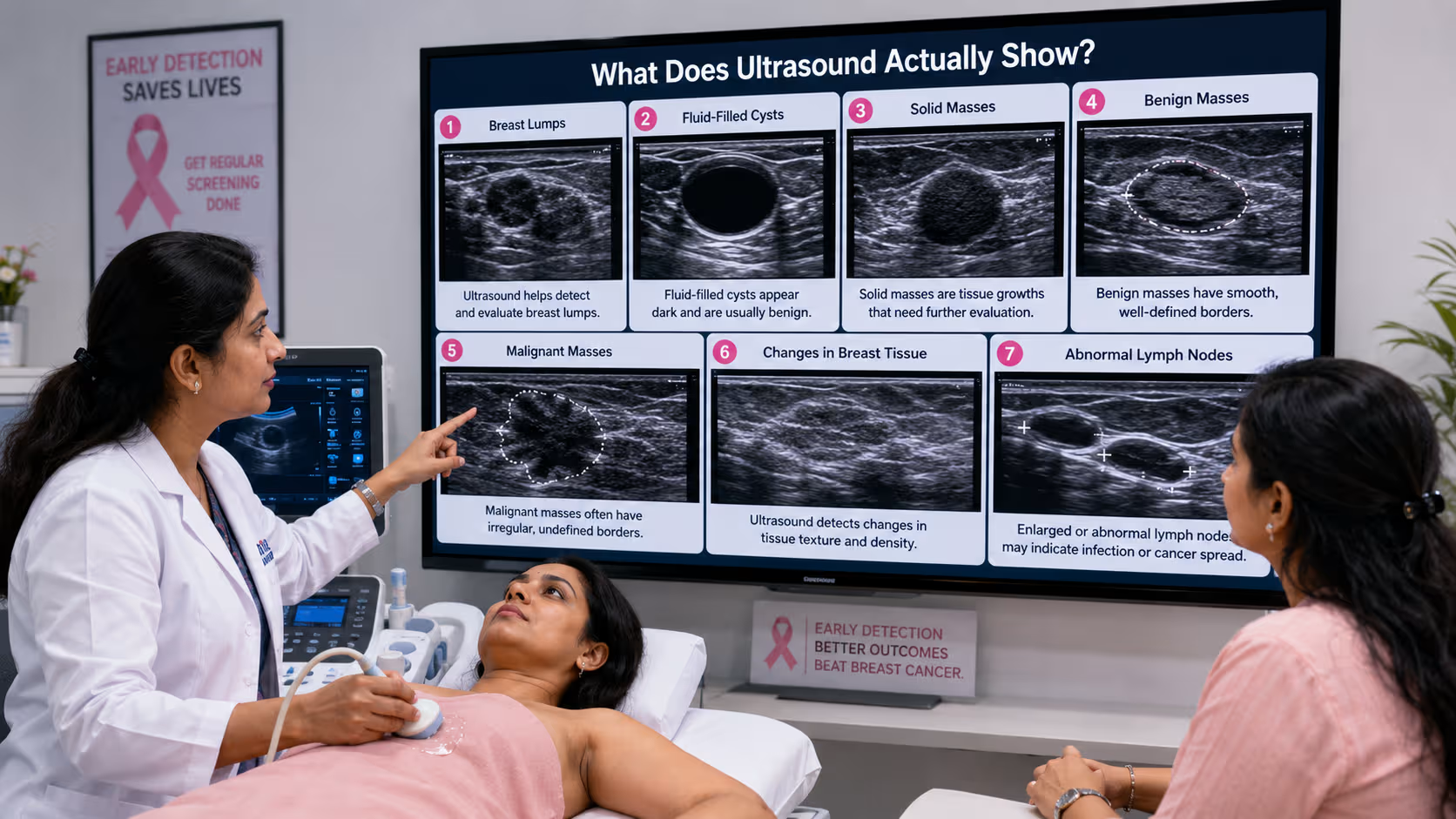
What Does Ultrasound Actually Show?
Ultrasound is exceptional at “characterizing” a finding. It helps doctors see through the “noise” of normal breast tissue.
Breast Lumps
When a woman feels a lump, an ultrasound is often the first step. It determines if the lump is something to worry about or a benign growth.
Fluid-Filled Cysts
This is where ultrasound excels. It can easily distinguish between a simple cyst (a sac filled with fluid, which is almost always non-cancerous) and a solid mass. Mammograms often cannot tell the difference between the two.
Solid Masses
If a mass is solid, the ultrasound helps the doctor look at its borders.
-
Benign masses usually have smooth, well-defined edges.
-
Malignant masses often have irregular, “spiculated” (star-shaped) edges or appear taller than they are wide.
Changes in Breast Tissue
Ultrasound can detect subtle distortions in the architecture of the breast tissue that might suggest an underlying pathology.
Abnormal Lymph Nodes
During a breast ultrasound, the specialist will often check the axilla (underarm) area. Enlarged or abnormally shaped lymph nodes can be an early sign that breast cancer is beginning to spread.
Can Ultrasound Detect Cancer Before Symptoms Appear?
The short answer is yes, but with caveats.
In women with dense breast tissue, ultrasound is often more sensitive than mammography. Dense tissue appears white on a mammogram, and so do tumors—making it like “looking for a snowball in a blizzard.” Ultrasound can see through that density.
Early Breast Abnormalities
Ultrasound can identify small tumors (less than 1cm) that are not yet palpable. However, it is not as good as mammography at detecting microcalcifications—tiny calcium deposits that are often the very first sign of ductal carcinoma in situ (DCIS), a non-invasive form of early cancer.
Limitations of Ultrasound
-
Operator Dependent: The accuracy of the test depends heavily on the skill of the person performing it.
-
False Positives: Ultrasound is very sensitive, meaning it finds many things that turn out to be harmless, which can lead to unnecessary biopsies and patient anxiety.
Quick Facts Box:
Accuracy: High for distinguishing cysts from solids.
Radiation: Zero.
Best For: Dense breasts and young women.
Main Drawback: Cannot see microcalcifications well.
Why Ultrasound Is So Helpful in Young Women?
Younger women (typically under 40) generally have higher breast density. Their breast tissue is packed with glandular and connective tissue and has less fat.
Dense Breast Tissue
As mentioned, mammography is less effective in dense breasts. For young women presenting with a lump, ultrasound is the primary diagnostic tool.
Breast Changes During Pregnancy
Hormonal shifts during pregnancy make breasts extremely dense and tender. Ultrasound is the safest and most effective way to evaluate any new lumps during this time without exposing the fetus to radiation.
Breastfeeding-Related Concerns
Conditions like mastitis (infection) or clogged ducts (galactoceles) are common during breastfeeding. Ultrasound can quickly differentiate an abscess that needs draining from a solid mass that needs a biopsy.
Evaluation of Breast Pain
While breast pain is rarely the only symptom of cancer, localized pain combined with a “thickening” of tissue is best evaluated via ultrasound to rule out underlying masses.
Ultrasound vs. Mammography: Which Is Better?
It isn’t a matter of which is “better,” but rather which is “right” for your specific breast type and age. Most experts, including Dr Farah Arshad, suggest they are complementary.
| Feature | Ultrasound | Mammography |
| Uses Radiation | No | Yes (Low dose) |
| Dense Breasts | Excellent visibility | Limited visibility |
| Detects Microcalcifications | Poor | Excellent |
| Detects Cysts | Excellent | Poor (shows as solid) |
| Screening Role | Supplemental | Primary (for 40+) |
| Comfort Level | High (Gel and probe) | Moderate (Compression) |
Detailed Explanation:
Mammography is the only screening tool proven to reduce breast cancer mortality in the general population because it catches microcalcifications. However, if a mammogram is “inconclusive” or shows “dense tissue,” an ultrasound is the essential next step to ensure nothing is hidden behind the dense glands.
What Happens If Ultrasound Finds Something Suspicious?
If an ultrasound reveals a solid mass with irregular borders or increased blood flow (detected via Doppler ultrasound), the medical team moves into the diagnostic phase.
BI-RADS Classification
Radiologists use the Breast Imaging-Reporting and Data System (BI-RADS) to score findings:
-
BI-RADS 1 & 2: Normal/Benign.
-
BI-RADS 3: Probably benign (usually requires a 6-month follow-up).
-
BI-RADS 4: Suspicious (biopsy is usually recommended).
-
BI-RADS 5: Highly suggestive of malignancy.
Additional Imaging
If the ultrasound is unclear, a Breast MRI might be ordered. MRI provides the most detailed images but is expensive and has a higher rate of false positives.
Needle Biopsy
If a mass looks suspicious, a Breast Cancer Specialist will perform an ultrasound-guided core needle biopsy. Using the ultrasound for “live” guidance, the doctor inserts a thin needle to take a small sample of the tissue for lab analysis. This is the only way to confirm if a mass is actually cancer.
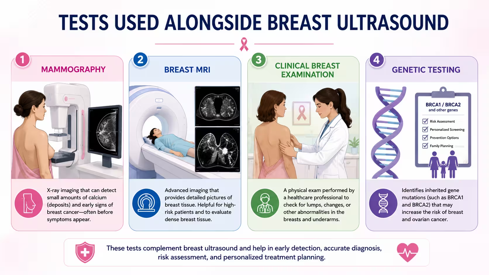
Other Tests Used Alongside Breast Ultrasound
Comprehensive breast care involves a “Multimodal Approach.”
-
Mammography: The baseline for women over 40.
-
Breast MRI: Used for high-risk patients (BRCA gene carriers) or to determine the extent of cancer after diagnosis.
-
Clinical Breast Examination: A physical exam by a specialist to feel for lumps or skin changes.
-
Genetic Testing: Recommended for women with a strong family history of breast or ovarian cancer to assess the risk of future occurrences.
Breast Cancer Treatment Options After Diagnosis
If an ultrasound and subsequent biopsy confirm cancer, the focus shifts to treatment. Modern medicine has made breast cancer highly treatable.
Surgery
-
Breast Conservation Surgery (Lumpectomy): Only the tumor and a small margin of healthy tissue are removed. This is often possible when ultrasound detects the cancer early.
-
Mastectomy: Removal of the entire breast tissue.
Radiation Therapy
High-energy beams are used to kill any remaining cancer cells after surgery.
Chemotherapy
Drugs used to shrink tumors or kill cancer cells that have spread.
Hormone Therapy
For cancers that are fueled by hormones (ER/PR positive), medications like Tamoxifen can block these hormones and prevent recurrence.
Targeted Therapy
Drugs like Herceptin target specific proteins (like HER2) on cancer cells, sparing healthy cells.
Immunotherapy and Newer Treatments
The landscape of cancer care is evolving rapidly, moving toward “Personalized Medicine.”
What Is Immunotherapy?
Immunotherapy helps your own immune system recognize and attack cancer cells. In breast cancer, it is particularly useful for Triple-Negative Breast Cancer (TNBC), which was historically harder to treat.
Recent Advances
-
Antibody-Drug Conjugates (ADCs): These are like “smart bombs” that deliver chemo directly to cancer cells.
-
Liquid Biopsies: Research is ongoing into blood tests that can detect cancer DNA before it even shows up on an ultrasound.
Is Breast Ultrasound Painful or Risky?
One of the reasons patients prefer ultrasound is its safety profile.
-
Safety: There are no known risks to ultrasound. It does not use radiation.
-
Comfort: The procedure involves a warm gel and a smooth probe. There is no painful compression of the breast tissue.
-
Pregnancy Safety: It is the primary tool for imaging during pregnancy and lactation.
Who Should Consider a Breast Ultrasound?
While not every woman needs an annual ultrasound, it is a vital tool for the following groups:
| Eligibility Group | Why Ultrasound? |
| Women with Lumps | To see if the lump is a cyst or a solid mass. |
| Dense Breast Tissue | To find tumors hidden by white glandular tissue. |
| Women Under 35 | Because their breasts are naturally denser and mammograms are less effective. |
| High-Risk Patients | As an add-on to mammograms and MRIs. |
| Abnormal Mammogram | To provide a “second look” at a specific area. |
Warning Signs of Breast Cancer You Should Never Ignore
While ultrasound can find cancer before symptoms appear, you must be vigilant about your own body. See a Breast Cancer Doctor in Lucknow immediately if you notice:
-
A New Lump: Especially one that feels hard, painless, and fixed in place.
-
Nipple Discharge: Any fluid other than breast milk, especially if it is bloody or clear/sticky.
-
Skin Dimpling: Skin that looks like an orange peel (peau d’orange).
-
Nipple Retraction: A nipple that has recently turned inward.
-
Breast Swelling: Redness or swelling of part of the breast even without a distinct lump.
-
Persistent Pain: While rare for cancer, any localized, non-cyclical pain should be checked.
Breast Cancer Awareness and Screening in Lucknow
In India, and specifically in cities like Lucknow, breast cancer is often diagnosed at a late stage due to a lack of awareness or fear of the diagnosis. However, clinical facilities in Lucknow are now equipped with world-class ultrasound and mammography technology.
Regular screening is the most powerful tool for the women of Lucknow. By opting for routine evaluations, women can ensure that if cancer does develop, it is caught at a stage where it is most curable. Experts like Dr Farah Arshad have been instrumental in promoting awareness and providing advanced diagnostic services to the community.
When Should You Consult a Breast Cancer Specialist?
You don’t need to wait for your annual check-up if something feels wrong. You should book a consultation with a Breast Cancer Specialist in Lucknow if:
-
You find a lump during a self-exam.
-
Your screening mammogram comes back as “Category 0” (meaning more imaging is needed).
-
You have a strong family history of cancer.
-
You are experiencing skin changes or nipple discharge.
For personalized care and expert guidance, Dr Farah Arshad is a recognized Breast Cancer Doctor in Lucknow, offering compassionate and comprehensive diagnostic and treatment services.
Final Thoughts
Can ultrasound detect breast cancer early? The answer is a resounding yes. While it is most effective when used in combination with mammography, ultrasound is an indispensable tool in the fight against breast cancer. It provides clarity, avoids radiation, and is a lifesaver for women with dense breast tissue.
Early detection is not just about the technology; it’s about the action you take. Regular screenings, self-exams, and consulting with the right specialists are the keys to a healthy future.
FAQs
1. Can ultrasound detect breast cancer early?
Yes, ultrasound can detect small, solid masses that are not yet palpable. It is particularly effective at finding early-stage cancers in women with dense breast tissue.
2. Is ultrasound better than mammography?
Neither is “better” overall. Mammography is better for finding calcium deposits (microcalcifications), while ultrasound is better for dense breasts and distinguishing cysts from solid tumors.
3. Can breast cancer be missed on ultrasound?
Like all tests, it is not 100% perfect. Some types of cancer, particularly those that don’t form a solid mass or those that look like normal tissue, can be missed. This is why using multiple methods (mammogram + ultrasound) is often recommended.
4. Who should get a breast ultrasound?
It is recommended for women with dense breasts, women under 35 with symptoms, pregnant women, and anyone who had an abnormal mammogram result.
5. Is breast ultrasound painful?
No. It is a painless procedure involving only a gel and a handheld probe. There is no compression involved.
6. How accurate is breast ultrasound?
It is highly accurate (over 90%) in distinguishing between a fluid-filled cyst and a solid mass. Its accuracy in diagnosing cancer increases when combined with clinical exams and mammography.
7. Can ultrasound distinguish a cyst from cancer?
Yes, this is one of the primary strengths of ultrasound. Cysts appear as black, clear circles, while solid masses (which could be cancer) appear as grey or white shaded areas.
8. What happens if a breast ultrasound is abnormal?
If the result is suspicious, your doctor will likely recommend a follow-up MRI or an ultrasound-guided biopsy to determine the nature of the mass.
9. Can young women use ultrasound for breast screening?
Yes, for women under 30-35, ultrasound is usually the first choice for breast imaging because of its safety and the density of young breast tissue.
10. When should I consult a breast cancer specialist?
Consult a specialist immediately if you find a lump, notice skin changes, have nipple discharge, or if your imaging report shows a BI-RADS score of 3 or higher.
CTA
Early breast cancer detection can significantly improve treatment success and long-term outcomes. If you notice a breast lump, persistent breast pain, nipple changes, or have concerns about your breast health, timely evaluation is essential.
For expert breast cancer screening, diagnosis, and personalized treatment guidance, consult Dr Farah Arshad, a trusted Breast Cancer Specialist in Lucknow and experienced Breast Cancer Doctor in Lucknow. Don’t wait—early detection saves lives. Book your consultation today.
